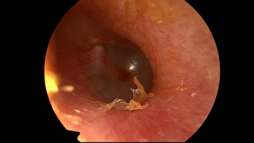
Pre-operative right otoendoscopic examination.
Right transcanal transpromontorial endoscopic approach for intralabyrinthine schwannoma - step by step
VIDEO


Intralabyrinthine schwannoma_Prof. Marchioni_Verona_Italy
PIEZOSURGERY® use
| RECOMMENDED INSERTS | |
Downloads
STEP BY STEP - PRE-OPERATIVE ASSESSMENT
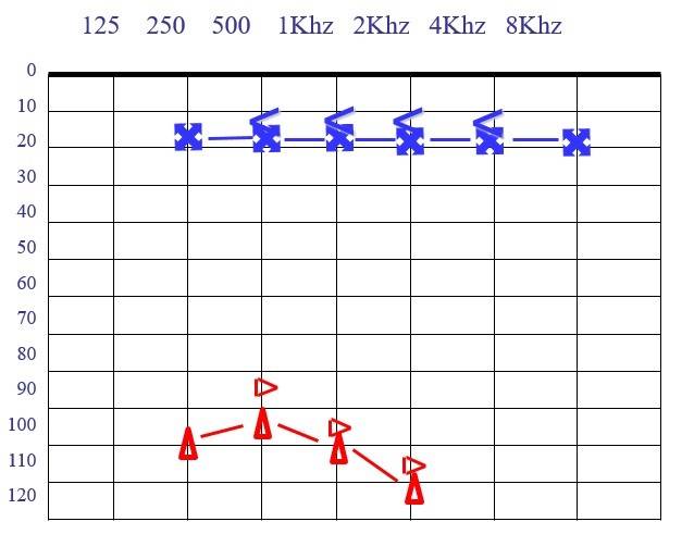
Pre-operative audiogram: normal left hearing and profound right sensorineural hearing loss.
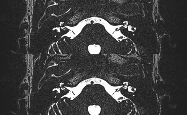
Axial view of CPA and brain MRI. Right intralabyrinthine schwannoma, involving the basal turn of the cochlea with extension to the fundus of the internal auditory canal.
STEP BY STEP - SURGICAL PROCEDURE

Right ear. Endoscopic view with a 0-degree endoscope; regular tympanic membrane. The skin of external auditory canal is injected.
A circumferential incision is made, using the monopolar with a needle tip.

The skin of external auditory canal is detached from the bone until reaching the anulus.
The tympanic membrane is elevated circumferentially.
The tympanic membrane is detached from the malleus and pulled inferiorly.
The skin of external auditory canal and the tympanic membrane are removed.
A large canalplasty and atticotomy are performed using a diamond burr and PIEZOSURGERY®.

The ossicular chain is exposed.
Removal of ossicular chain and chorda tympani in order to expose the medial wall of tympanic cavity.

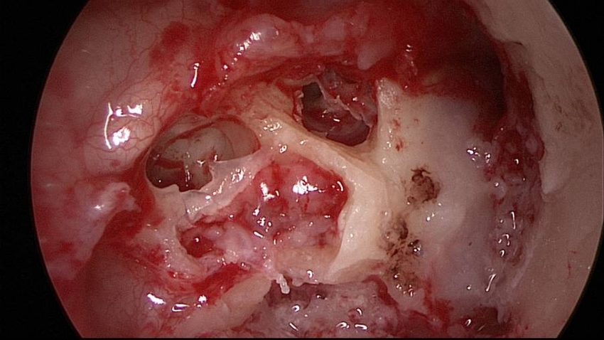
Endoscopic view of the round window chamber.
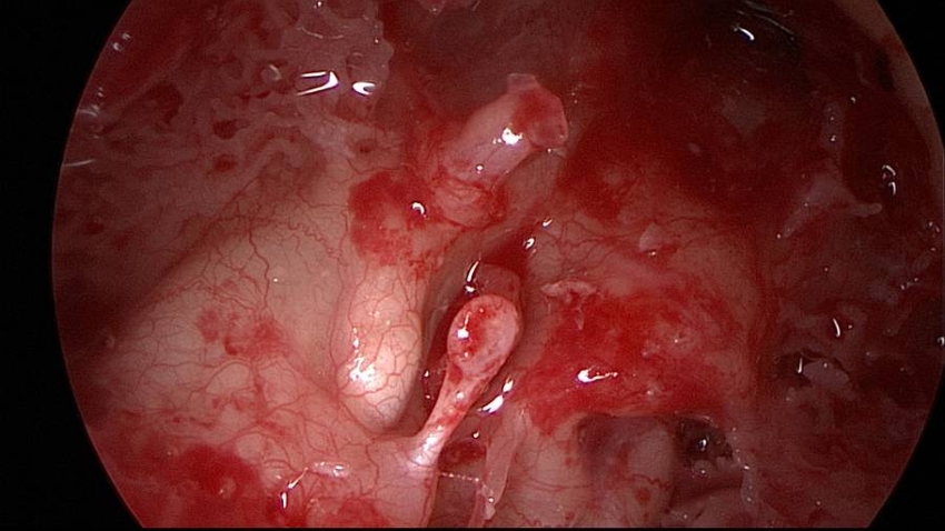
Endoscopic view of the medial wall of tympanic cavity after canalplasty and ossicular chain removal.
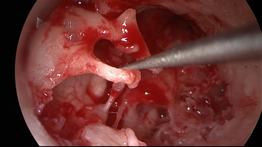
Stapes removal.
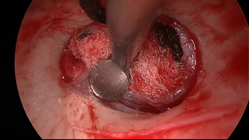
Promontory region is drilled using PIEZOSURGERY®, in order to reach the schwannoma.

Promontory region is drilled using PIEZOSURGERY®, in order to reach the schwannoma.
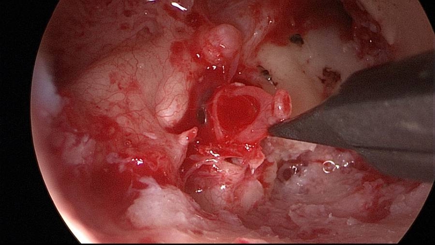
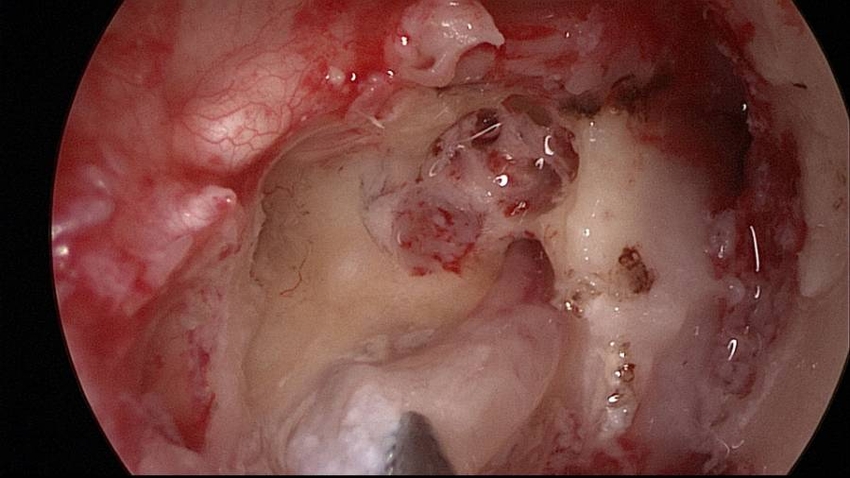
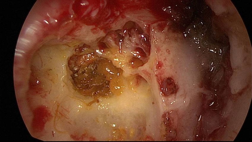
The vestibule is opened, the tensor tympani muscle is pulled forward and the cochlea is opened exposing the schwannoma, filling the basal turn.

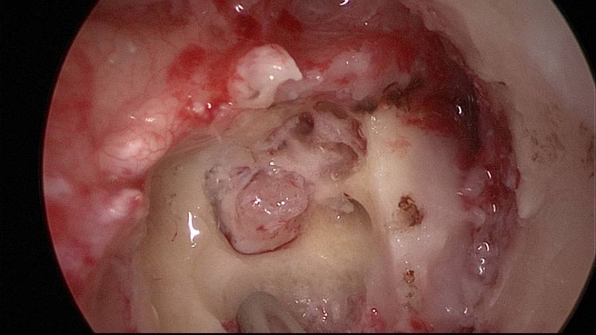
Further drilling with PIEZOSURGERY® is performed in order to exposed the intracameatal extension of the schwannoma. Cerebrospinal fluid leak occur, after opening the fundus of internal auditory canal.
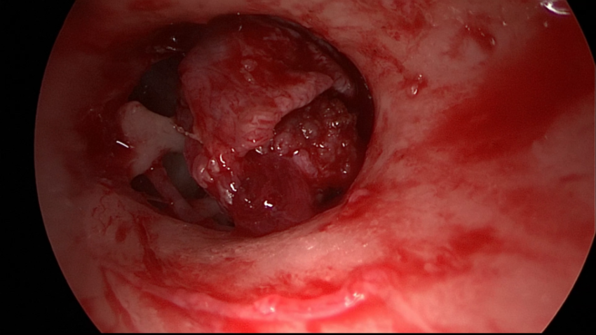
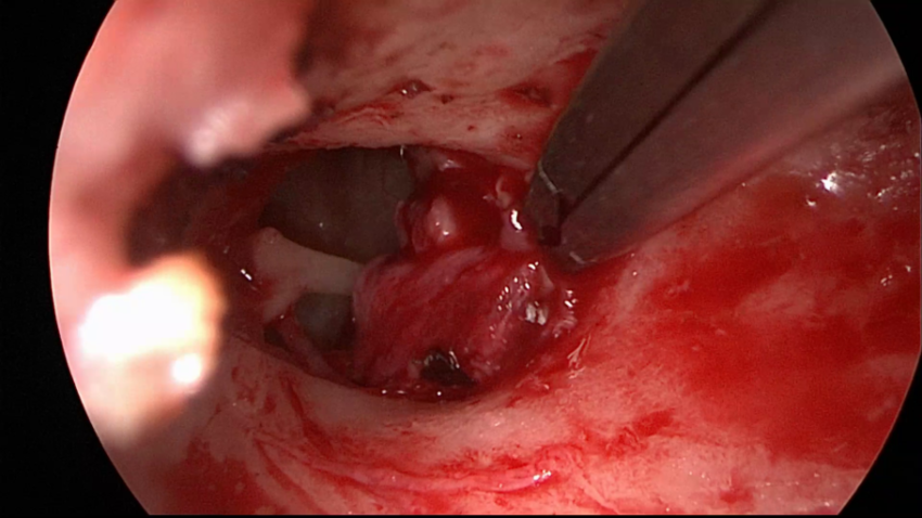
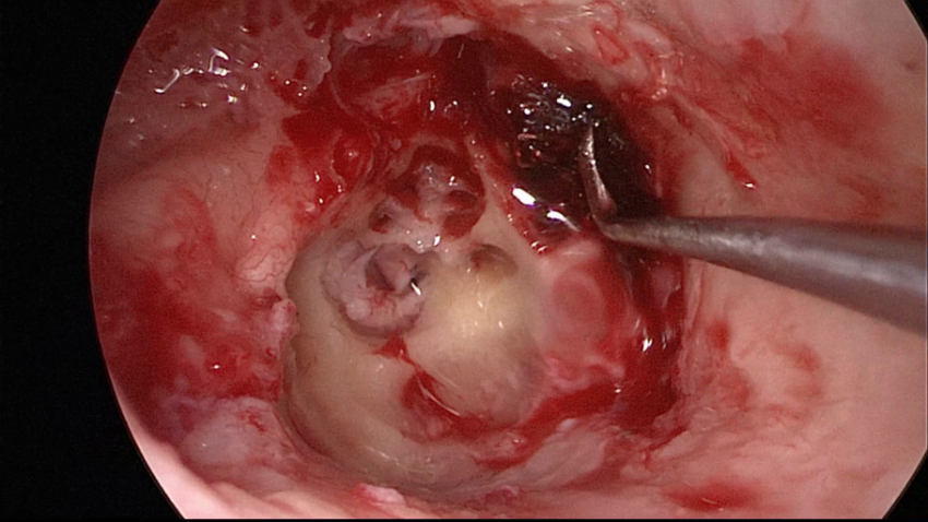
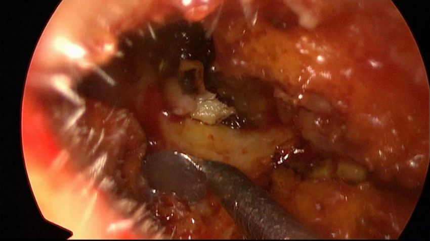
The schwannoma into the basal turn of the cochlea is removed.
The intrameatal portion of the schwannoma is also removed.

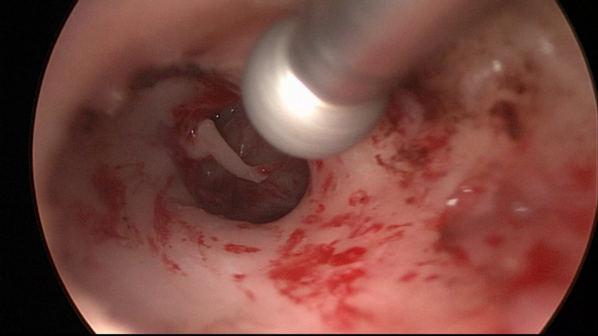
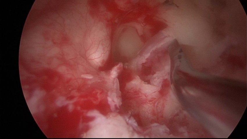
Final surgical cavity after schwannoma removal. The openings of the cochlea and the internal auditory canal are showed.
The nerves inside the internal auditory canal are exposed: the cochlear nerve to the modiolus, the inferior and superior vestibular nerves, the facial nerve in a deeper posisition.
The Eustachian tube is obliterate with tensor tympani muscle pulled forward and hemostat absorbable material.
The surgical cavity is obliterate with muscle, fibrin glue and hemostat absorbable material.
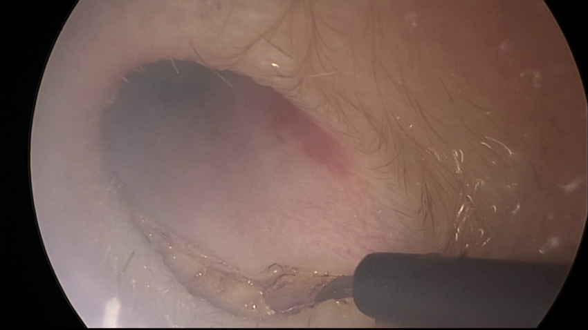
Eversion of the external auditory canal skin and blind sac closure of the canal are performed.