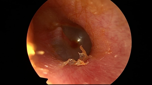
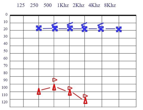
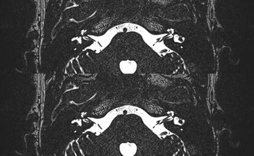
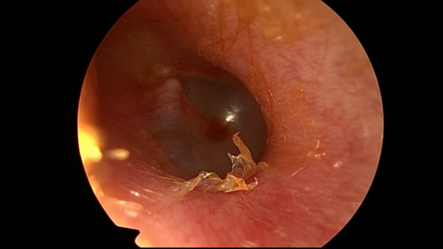
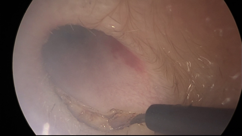
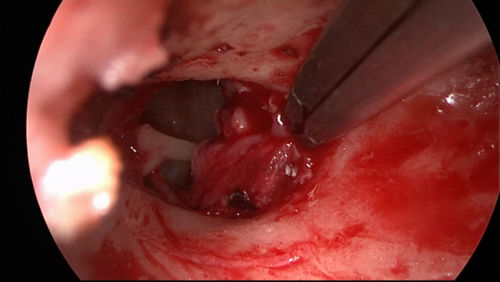
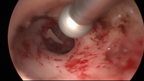
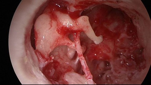
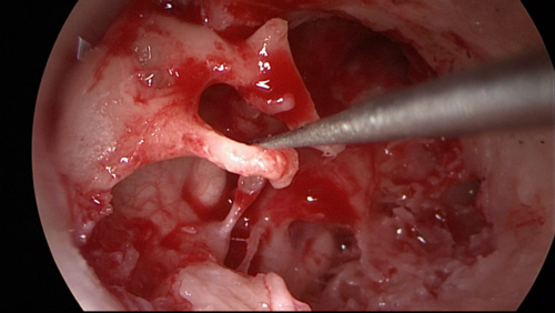
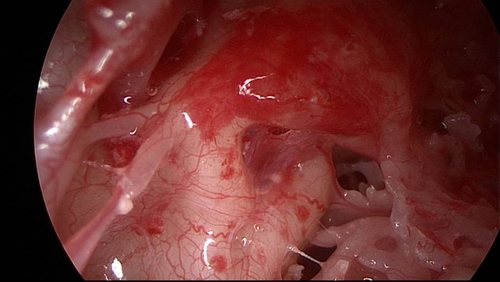
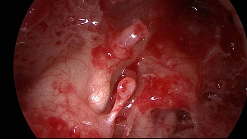
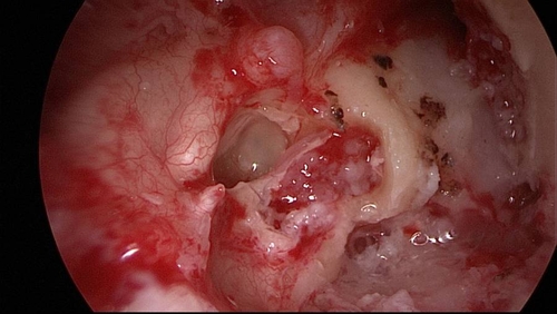
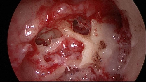
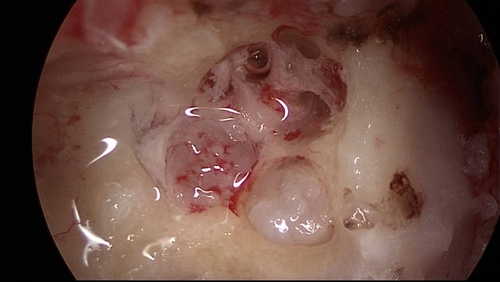
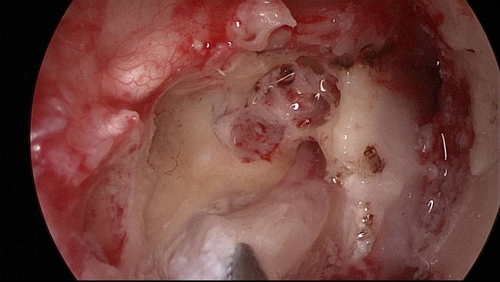
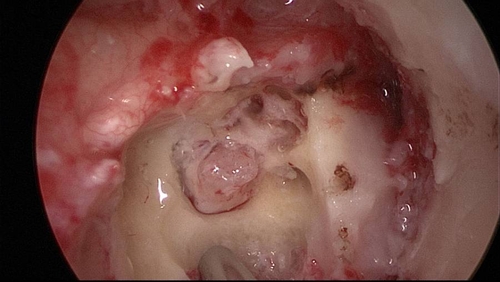
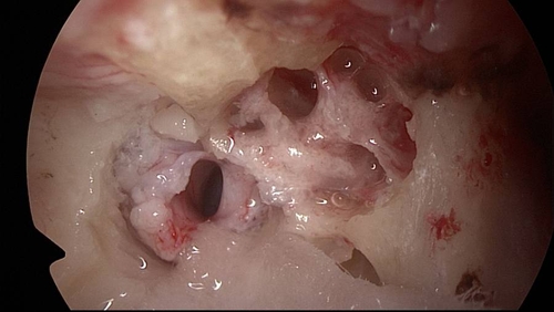
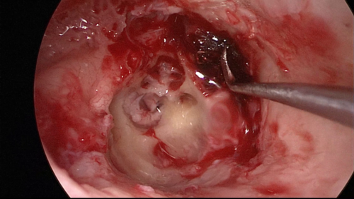
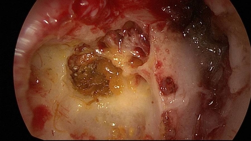
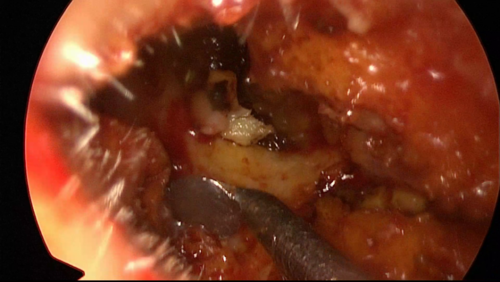
Pre-operative right otoendoscopic examination.
Right transcanal transpromontorial endoscopic approach for intralabyrinthine schwannoma
PIEZOSURGERY® Einsatz
| EMPFOHLENE INSTRUMENTE | |
Downloads
STEP BY STEP - VOROPERATIVE BEURTEILUNG
STEP BY STEP - CHIRURGISCHES VORGEHEN
Vorgehensweise Schritt für Schritt beschrieben
- The skin of external auditory canal is injected, then a circumferential incision is made, using the monopolar with a needle tip.
- The skin of external auditory canal is detached from the bone until reaching the annulus.
- The skin of external auditory canal and the tympanic membrane are removed.
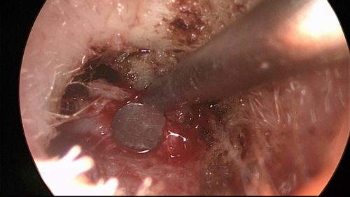
- A large canalplasty and atticotomy are performed using a diamond burr and/or Piezosurgery.
Notice how the Piezosurgery allows a constant cleaning of the surgical field during this phase, thanks to its continuous washing. On the other hand, the traditional burr burns bone tissue and produces bony dust, obscuring the endoscopic view.
- The ossicular chain is exposed and removed in order to expose the medial wall of tympanic cavity.
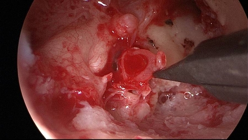
- Promontory region is removed using Piezosurgery, in order to reach the schwannoma into the cochlea.
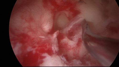
- Further drilling with Piezosurgery is performed in order to exposed the intrameatal extension of the schwannoma.
During the opening of the fundus of the internal auditory canal, it is preferred to use the Piezosurgery because it allows a precise drilling preserving the nervous structures located into the canal.
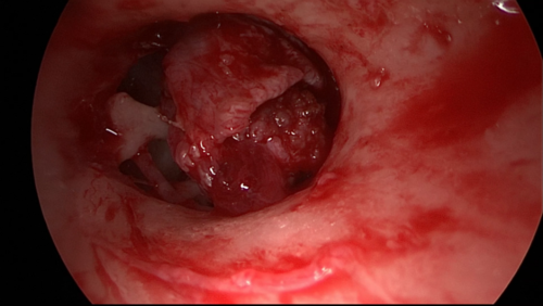
- The schwannoma into the basal turn of the cochlea with the intrameatal portion is removed.
- The openings of the cochlea and the nerves inside the internal auditory canal are exposed.
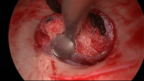
- The Eustachian tube is obliterate with tensor tympani muscle pulled forward and hemostat absorbable material.
- The surgical cavity is obliterate with muscle, fibrin glue and hemostat absorbable material.
- Eversion of the external auditory canal skin and blind sac closure of the canal are performed.


